J. Wearing1,2,*, M. Stokes3,4, R.A. de Bie1, E.D. de Bruin5,6
1. Department of Epidemiology, Faculty of Health, Medicine and Life Sciences, School for Public Health and Primary Care, Maastricht University, Maastricht, The Netherlands; 2. Adullam Spital und Pflegezentren, Basel, Switzerland; 3. School of Health Sciences, University of Southampton, Southampton, United Kingdom; 4. Centre for Sport, Exercise and Osteoarthritis Research Versus Arthritis, Nottingham, United Kingdom; 5. Institute of Human Movement Sciences and Sport (IBWS) ETH, Department of Health Sciences and Technology, ETH Zurich, Zürich, Switzerland; 6. Division of Physiotherapy, Department of Neurobiology, Care Sciences and Society, Karolinska Institute, Stockholm, Sweden. Corresponding author: Julia Wearing, Adullam Spital und Pflegezentren, Basel, Switzerland, E-mail: j.wearing@bluewin.ch, Phone: +41 61 2669799
Jour Nursing Home Res 2020;6:120-126
Published online December 16, 2020, http://dx.doi.org/10.14283/jnhrs.2020.30
Abstract
Background: Age-related neurological and muscular changes lead to low strength and function in older people. The proportional contribution of these changes to strength decline alters with increasing age. However, it is not clear how the muscle changes in older people which have excessive loss of strength due to multimorbidity and inactivity. Unlike community-living older adults, intramuscular alterations are rarely evaluated in nursing-home residents despite their potential importance for specific sarcopenia assessment and guiding interventions to improve strength. Objectives: To explore potential relationships between muscle strength, muscle quantity, contractile quality and physical activity in older nursing-home residents. Design: A cross-sectional proof-of-concept study. Setting: A nursing-home in Switzerland. Participants: 24 nursing-home residents, median age (range) 86.5 (68-103) years. Measurements: Sonographic measures of muscle thickness and echotexture were used as surrogate measures of muscle quantity and contractile quality of the quadriceps muscle. The relationship between sonographic measures and isometric strength of the knee extensors, gait speed and physical activity was evaluated using Pearson’s and Spearman’s correlation coefficients. A subgroup analysis of low (n=12) and normal (n=12) physical activity, based on energy expenditure cut off values of 383 kcal/week for men and 270 for women, was also undertaken. Results: In nursing home residents with normal physical activity, muscle quality positively correlated with knee extensor strength (r=0.727, p=.007) and gait speed (r=0.588, p=.044) while muscle thickness was not (p=0.966 and p=.564 respectively). There was no correlation among variables for n=24 or the subgroup with low physical activity. Conclusions: The results provide proof of concept that poor muscle quality is associated with low strength in older nursing-home residents that are physically active. Ultrasound derived muscle quality assessment has potential to detect activity-related muscle differences in old age, associated with sarcopenia, and may be more appropriate than muscle thickness measures.
Key words: Knee extensor strength, muscle quality, heterogeneity, nursing-home residents, physical activity.
Introduction
Age-related neurological and muscular changes lead to low strength in older people with an increased risk of functional decline (1). Residents of nursing homes, in particular, often have excessive muscle weakness (2) due to multiple chronic diseases, inactivity and malnutrition (1). To prevent adverse health outcomes, assessment of muscle disorders, and preservation or improvement in muscle strength are essential (3).
For several decades, the age-related muscle disorder sarcopenia has been mainly attributed to a reduction in muscle mass (1). Recently, however, it has been shown that the relationship between strength and muscle mass is inconsistent in older people and that strength decreases to a higher extent than mass with aging (4). Explanatory models have now highlighted the potential role of muscle quality and neural function, in addition to muscle mass, as key determinants of muscle strength (4). Muscle quality has been shown to decrease with age but is suspected to change more due to muscle disuse, which often accompanies aging (5). In the clinical context of strength, muscle quality refers to contractile characteristics of muscle fibres (5) and can be determined e.g. by the ratio of muscle strength per body mass or muscle imaging techniques (6). The quality of muscular contraction is thought to be diminished by multiple factors (5), including the accumulation of intramuscular connective and adipose (non-contractile) tissue (7), muscle fibre necrosis and inflammation that change the density and heterogeneity of the muscle structure (8).
Evaluation of intramuscular non-contractile tissue has been recently applied in research that focuses on age-related changes in muscle as well as in patients with muscle impairments such as muscular dystrophy (5, 9). While Magnetic Resonance Imaging has been widely used to quantify morphologic abnormalities in muscles, B-mode ultrasound imaging has been shown to be a reliable alternative in clinical settings, complementing functional measures of muscles and demonstrating disease progression by changes in thickness and echo intensity (9). Clinical trials evaluating healthy older, community-living people have shown that strength consistently declines along with altered muscle quality, however its relation to muscle quantity is controversial (10-12). Moreover, muscle quality has been shown to be positively related to gait performance (12, 13). Despite the high informative value of ultrasound measures in detecting muscle changes in healthy elderly, there is a lack of studies using ultrasound in older (≥ 65 years), particularly in oldest-old (≥ 85 years), comorbid people (14).
Only a few studies to date have evaluated muscle characteristics in nursing-home residents. As muscle strength decreases with age, particularly rapidly after the age of 80 years (15), the proportional contribution of muscular and neurological changes leading to strength decline also changes (4, 15). Particularly in people with a very low muscle condition such as in nursing-home residents (1, 2, 16), the muscle might show a specific pattern of biological changes. Moreover, as the evaluation of muscle function by assessments typically used in older adults is limited in people with very low muscle condition due to physical restrictions (17), evaluation of ultrasound-derived muscle changes might be a valuable alternative for this cohort. Therefore, morphologic changes in muscle, and relationships between strength, function and physical activity in this population merit separate attention to improve clinical assessment of sarcopenia in this population. So far, it has been shown in computer-tomographical scans that cross-sectional area of the quadriceps femoris was related to knee extensor strength and gait speed in frail, older nursing-home residents (18). However, whether there is a relationship between ultrasound measures of muscle morphology and strength of the lower extremities in nursing-home residents has not been explored to date (7).
The objectives of this study were 1. To explore potential relationships between sonographic measures of muscle quantity, quality and strength of the knee extensors in older nursing-home residents, specifically including oldest-old people, and 2. To examine the relationships of knee extensor characteristics and gait speed within subgroups based on physical activity level.
Methods
Study design
An observational, cross-sectional proof-of-concept (19) study design was used to explore the potential relationships between muscle strength, sonographically measured thickness, indices of quality, and physical function in a convenience sample of older, comorbid residents of a nursing-home in Switzerland, including oldest-old people (≥ 85 years).
Participants recruitment
Residents were included if they were able to walk, with or without the aid of a walking device, and cognitively able to understand study content. Exclusion criteria were a) severe impairment in decision making (Cognitive performance scale > 4 points) (20), b) acute illness, c) a history of acute lower limb pathology within the last 6 months (fracture and/or surgery) and d) skin disorders involving the anterior thigh.
All participants provided informed consent following a verbal and written explanation of the study procedures, which complied with the principles of the Declaration of Helsinki for ethical research in humans. The study received approval from the local ethics committee (Ethikkommission Nordwest- und Zentralschweiz (EKNZ), project-ID 2017-00839).
Data collection
Participants demographics
Age, height and body mass of the participants and number of chronic diseases that may affect muscle characteristics (metabolic, musculoskeletal, neurological, psychiatric, respiratory disease, renal insufficiency and cancer) were extracted from nursing assessments and medical history.
Muscle strength, muscle morphology, physical function and physical activity were examined by a physiotherapist trained and experienced in musculoskeletal assessments.
Muscle strength
Maximal isometric strength of the knee extensor was measured with a hand-held dynamometer (Microfet®, CompuFET, Hoggan Health Industries, Biometrics Europe). For measurements, participants were seated on a plinth, with their back and thighs fully supported, knee positioned at 90° and lower leg hanging freely. The curved transducer pad of the dynamometer was positioned at 80% of the length of the tibia. Participants were requested to push against the dynamometer as hard as possible for 3 seconds.
The peak force measured during two trials was recorded in kg. Relative strength was calculated by normalizing peak force to body mass. Isometric muscle strength determined by hand-held dynamometry in older adults has been shown to be comparable to strength values measured with the gold standard method of isokinetic strength testing (21) with a high test-retest reliability (ICC 0.90-0.98) (22).
Muscle morphology
Real-time, B-mode ultrasonography (Nemio MX Type SSA-590A, Toshiba, Japan) with a 12-MHz linear transducer array (45 mm footprint) was used to obtain two transverse scans of the rectus femoris and vastus intermedius muscles at a site two-thirds of the distance between the antero-superior iliac spine and the superior pole of the patella. Standardized sonographic settings were used for all participants and images were acquired using a uniform protocol developed for older adults (23). Ultrasound images were post-processed using a MATLAB code (MathWorks®, Massachusetts, USA). Muscle thickness was calculated as the distance between the fascial layers that distinguish muscles from the subcutaneous fat layer, and muscles from bone. Thickness values were expressed as a percentage of total thigh thickness to account for individual body composition (23). Sonographic measures of quadriceps thickness have been previously shown to be highly correlated (r = 0.98) with gold standard measures obtained from Magnetic Resonance Imaging (14), and have a reported intra-rater reliability of ICC 0.88-.099 in older people (8, 23).
Muscle echotexture was characterized by echo intensity and heterogeneity, determined over a rectangular region of interest. Echo intensity was estimated by calculating the mean grayscale value in unspecified units (UU), ranging between 0–255. High values reflected hyperechoic tissue which reportedly reflects the accumulation of non-contractile tissue (9), muscle fibre necrosis and inflammation (8). Tissue heterogeneity was estimated by calculating the standard deviation (SD) of grayscale value (24). Low SD reflected low heterogeneity which indicates homogenous tissue structure and evenly distributed non-contractile tissue within muscle (24). Homogeneous muscle tissue patterns have been shown to be positively associated with muscle disorders (24), and age-related strength differences (25). Quantification of tissue heterogeneity has been demonstrated to be sensitive in detecting neuromuscular disorders including myopathy and muscle dystrophy (26).
Physical function
Preferred gait speed was evaluated over a 4m distance. This method is commonly used for evaluation of functional performance in older adults and shows excellent test-retest reliability in older people with comorbidities (17).
Physical activity
Physical activity was evaluated using the German Physical Activity 50+ questionnaire (27), which measures the duration of various household and leisure time activities and allows for estimation of energy expenditure based on the Compendium for Physical Activities (28). Its performance characteristics have been reported elsewhere (27). Energy expenditure was reported in kcal/week and dichotomized into low- and normal physical activity based on cut off values of 383 and 270 kcal/week for men and women, respectively (29). For evaluation of physical activity, participants as well as nurses responsible for their care, who closely observe the nursing-home residents 24h/7d were interviewed, to increase precision of the responses.
Statistical analysis
Normal distribution of the data was evaluated using the Shapiro-Wilk test. Normally distributed data have been reported as mean (SD), not normally distributed data as median (range). For normally distributed data, Pearson’s correlation coefficient was used to evaluate correlations between measures of muscle strength, muscle morphology, physical performance and physical activity, and to analyze relationships between knee extensor characteristics within the physical activity subgroups. For not normally distributed data, Spearman’s correlation coefficient was used accordingly.
Results
Descriptive Analysis
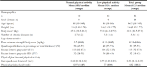
The study sample included n=24 participants (18 women), median age (range) 86.5 (68-103) years, with n=12 participants in each physical activity-subgroup (Table 1).
†data not normally distributed
Correlation analysis (n=24)
Knee extensor strength normalized to body mass was not significantly correlated to sex or measures of muscle morphology, physical function or physical activity (Table 2).
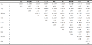
Table 2
Correlations among demographic parameters, quadriceps muscle strength and morphology, physical function
and physical activity (n= 24)
CD = number of chronic diseases, KES = knee extensor strength normalized to body mass, QT = quadriceps thickness in relation to total thickness, RG = Rectus grayscale, RGSD = rectus grayscale standard deviation, GS = gait speed, PA = physical activity, Significance p<.05; †correlations determined by Spearman’s Correlation Coefficient; Body mass was excluded from the correlation analysis as muscle strength has been normalized to mass.
Correlations within physical activity subgroups (n=12 each)
To determine if a higher activity level, mainly achieved by walks in the garden and outside the nursing-home, is important to determine relationships of morphological measures and strength of the quadriceps, the sample was divided into two subgroups categorized by the physical activity cut-off value of 270kcal for women and 383kcal for men. The cut off value determines whether a person is at risk for frailty (29). Both subgroups included n=12 participants with an energy expenditure of mean (SD) 68 kcal/week (100) in the slow group and 1297 kcal/week (1445) in the group with normal physical activity.
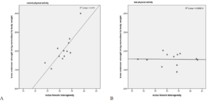
Within the subgroup with normal physical activity, knee extensor strength was positively correlated with tissue heterogeneity (r = 0.727, p = .007) but not with muscle thickness (r = 0.014, p = .966). Similarly, muscle tissue heterogeneity was positively correlated with gait speed (r = 0.588, p = .044). However, within the subgroup of people with low physical activity, there were no significant correlations among knee extensor strength normalized to body mass, comorbidities, muscle thickness and muscle echo intensity and heterogeneity, or gait speed (Table 3).
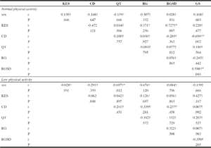
Table 3
Correlation analysis between muscle characteristics of the knee extensor and gait speed within subgroups of physical activity
CD = number of chronic diseases, KES = knee extensor strength normalized to body mass, QT = quadriceps thickness in relation to total thickness, RG = Rectus grayscale, RGSD = rectus grayscale standard deviation, GS = gait speed, PA = physical activity; †correlations determined by the Spearman’s correlation coefficient; *significance, p<.05
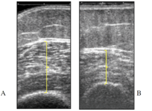
Correlations of rectus femoris tissue heterogeneity with knee extensor strength, in those with normal and low physical activity are shown in Figure 1. Figure 2 presents two ultrasound images with different muscle tissue heterogeneity.
Scatterplot of rectus femoris tissue heterogeneity and knee extensor strength normalized to body mass in those with normal physical activity (A) and low physical activity (B).
Two ultrasound images with similar grayscale value of muscle tissue (region indicated by yellow line) but different in grayscale standard deviation (SD): (A) higher grayscale SD (heterogeneous tissue) of a participant with normal physical activity level, (B) lower grayscale SD (homogenous tissue) of a participant with low physical activity level; the length of the yellow arrow defines the muscle thickness between subcutaneous tissue above it and bone below.
Discussion
This study explored potential relationships among ultrasound derived indices of muscle quality characteristics, muscle function (gait speed) and physical activity in older, multimorbid nursing-home residents. This proof-of-concept study targeting muscle aging mechanisms showed that ultrasound assessments explored the relationship between muscle quality, strength of the knee extensors and gait speed in older (68-103 years) nursing-home residents with normal physical activity. Ultrasound imaging seems to be a useful measure for detecting physical activity-related muscle differences in this cohort, contributing to comprehensive sarcopenia assessment. It can be useful to detect differences in muscle quality associated with limitations in muscle function in people not capable of performing functional measures of strength.
While knee extensor strength values in the current study are comparable to strength values reported for nursing-home residents of other European countries (18), strength and muscle morphology were not related to age. Although muscle characteristics change over the lifespan, differences within a group of older, comorbid people might be more related to disuse than to age per se (1). The relationship between knee extensor strength, muscle thickness and muscle morphology differed depending on the physical activity level in this sample of participants. The cut-off value for low- and normal-physical activity (279/383 kcal per week) in the current study is equivalent to the energy expended by a 50-kg body mass woman whose only activity is to walk at a comfortable speed for 20 minutes a day, 5 times a week. Physical activity at this level reflects very little activity even for nursing home residents and is considered to be indicative of high disability (16).
In elderly residents with normal physical activity, tissue heterogeneity, a measure of muscle quality (6, 25), correlated with knee extensor strength but not thickness. This finding is consistent with previous literature in community-living elderly (10-13). Muscle quality might, therefore, be a more appropriate indicator of age-related changes in muscle strength than muscle thickness. In the present study, muscle heterogeneity was not only related to strength in the subgroup with normal physical activity but also to gait speed, an important measure of functional status (1). Even if a causal relationship cannot be established by these data, they indicate mutual impact between these factors. It is possible that minimizing the accumulation of non-contractile tissue in muscle through physical activity interventions (5) could also effectively improve non-contractile tissue dispersion and positively affect gait. Further research evaluating the impact of physical activity on muscle heterogeneity could verify this suggestion.
Within the low physical activity subgroup, no significant relationships were observed between knee extensor strength and common sonographic measures of muscle morphology (muscle thickness and echo intensity). This finding cannot be compared to the literature as, to our knowledge, this is the first study to evaluate those relationships in nursing-home residents concerning physical activity. One potential reason for the present results might be that once muscle strength drops below a certain threshold, morphological interactions are less apparent. Alternatively, it is possible that the sonographic approach in the present study is not sufficiently sensitive to detect physical activity-related differences in muscle of highly reduced condition. The detection of catabolic biomarkers in blood samples could potentially give more detailed information about muscular changes (5). Another reason might be that neurological components of strength were not considered in this study as an explanatory factor for low strength. However, echotexture analysis of ultrasound images provides important information in the context of age-related muscle disorder even in older, comorbid nursing-home residents.
The present findings are indicative of the following hypotheses: 1. Muscle quality contributes to age-related decrease in strength in nursing-home residents. Further research is needed to verify this hypothesis and should also focus on the neurological determinant of muscle strength. 2. Grayscale SD evaluated by echo intensity of ultrasound images could be used to detect differences in age-related muscle quality. The accordance of different methods detecting heterogeneity in muscle structure would have to be evaluated.
Limitations
One potential limitation of this study is that tissue heterogeneity is likely to vary within a given muscle and may be dependent on subcutaneous tissue thickness (25). While this study adopted a standardized region of interest for estimating tissue heterogeneity, it should be recognized that calculation of parameters at the midpoint of the muscle, without taking subcutaneous tissue into consideration, may not be reflective of the whole muscle unit. The findings are, however, consistent with other studies in which tissue heterogeneity has been shown to be altered in neuromuscular disease and age-related muscle wasting (24, 25). Secondly, we cannot be certain that the isometric strength measurements involved maximal effort of the participants. Discrepancies between voluntary muscle contraction und maximal possible contraction could have been detected by electrical muscle stimulation using the interpolated twitch technique (30) but this is not feasible outside of laboratory settings. Therefore, the assessment of maximal isometric voluntary contraction is an appropriate alternative as it has been shown to be reliable in strength detection of older adults in clinical settings (22). Although further studies are needed to verify the results, the findings of the present study provide new insights into muscle characteristics in older, including oldest-old, institutionalized people.
Conclusions
The present results provide proof-of-concept that muscle tissue heterogeneity, a sonographically measured index of muscle quality, is positively related to knee extensor strength and gait speed, in older nursing-home residents with normal physical activity level. The findings indicate that the assessment of muscle quality from ultrasound images might be a more appropriate method to detect age-related differences in muscle than the evaluation of muscle thickness, at least in older people with normal physical activity. Given the sample size of the current study (n = 24), assessment of ultrasound-derived muscle morphology in a larger population sample is warranted. Inclusion of these assessments may be recommended as part of a comprehensive assessment of sarcopenia and for monitoring the outcome of preventative interventions designed to improve muscle strength and function in older individuals.
Funding: The authors received no specific funding for this work.
Acknowledgments: We would like to thank the nursing-home residents for their participation, Dr. Hans-Jörg Ledermann for his valuable suggestions during the planning of this research work, and the nursing staff for their help in recruitment and data collection. We acknowledge Adullam Spital und Pflegezentrum Basel for providing equipment.
Conflict of interest: Julia Wearing declares that she has no competing interests. Maria Stokes declares that she has no competing interests. Rob A. de Bie declares that he has no competing interests. Eling D. de Bruin declares that he has no competing interests.
References
1. Cruz-Jentoft, A.J. and A.A. Sayer, Sarcopenia. Lancet, 2019. 393(10191): p. 2636-2646.
2. Shen, Y., et al., Prevalence and Associated Factors of Sarcopenia in Nursing Home Residents: A Systematic Review and Meta-analysis. J Am Med Dir Assoc, 2019. 20(1): p. 5-13.
3. Fragala, M.S., et al., Resistance Training for Older Adults: Position Statement From the National Strength and Conditioning Association. J Strength Cond Res, 2019. 33(8): p. 2019-2052.
4. Manini, T.M. and B.C. Clark, Dynapenia and aging: an update. J Gerontol A Biol Sci Med Sci, 2012. 67(1): p. 28-40.
5. Fragala, M.S., A.M. Kenny, and G.A. Kuchel, Muscle quality in aging: a multi-dimensional approach to muscle functioning with applications for treatment. Sports Med, 2015. 45(5): p. 641-58.
6. Correa-de-Araujo, R., et al., The Need for Standardized Assessment of Muscle Quality in Skeletal Muscle Function Deficit and Other Aging-Related Muscle Dysfunctions: A Symposium Report. Front Physiol, 2017. 8: p. 87.
7. Addison, O., et al., Intermuscular fat: a review of the consequences and causes. Int J Endocrinol, 2014. 2014: p. 309570.
8. Mayans, D., M.S. Cartwright, and F.O. Walker, Neuromuscular ultrasonography: quantifying muscle and nerve measurements. Phys Med Rehabil Clin N Am, 2012. 23(1): p. 133-48, xii.
9. Simon, N.G., Y.I. Noto, and C.M. Zaidman, Skeletal muscle imaging in neuromuscular disease. J Clin Neurosci, 2016. 33: p. 1-10.
10. Fukumoto, Y., et al., Skeletal muscle quality assessed from echo intensity is associated with muscle strength of middle-aged and elderly persons. Eur J Appl Physiol, 2012. 112(4): p. 1519-25.
11. Watanabe, Y., et al., Echo intensity obtained from ultrasonography images reflecting muscle strength in elderly men. Clin Interv Aging, 2013. 8: p. 993-8.
12. Rech, A., et al., Echo intensity is negatively associated with functional capacity in older women. Age (Dordr), 2014. 36(5): p. 9708.
13. Akazawa, N., et al., Relationships between intramuscular fat, muscle strength and gait independence in older women: A cross-sectional study. Geriatr Gerontol Int, 2017. 17(10): p. 1683-1688.
14. Ticinesi, A., et al., Muscle Ultrasound and Sarcopenia in Older Individuals: A Clinical Perspective. J Am Med Dir Assoc, 2017. 18(4): p. 290-300.
15. Hunter, S.K., H.M. Pereira, and K.G. Keenan, The aging neuromuscular system and motor performance. J Appl Physiol (1985), 2016. 121(4): p. 982-995.
16. Buckinx, F., et al., Relationship between ambulatory physical activity assessed by activity trackers and physical frailty among nursing home residents. Gait Posture, 2017. 54: p. 56-61.
17. Beaudart, C., et al., Assessment of Muscle Function and Physical Performance in Daily Clinical Practice : A position paper endorsed by the European Society for Clinical and Economic Aspects of Osteoporosis, Osteoarthritis and Musculoskeletal Diseases (ESCEO). Calcif Tissue Int, 2019. 105(1): p. 1-14.
18. Casas-Herrero, A., et al., Functional capacity, muscle fat infiltration, power output, and cognitive impairment in institutionalized frail oldest old. Rejuvenation Res, 2013. 16(5): p. 396-403.
19. Justice, J., et al., Frameworks for Proof-of-Concept Clinical Trials of Interventions That Target Fundamental Aging Processes. J Gerontol A Biol Sci Med Sci, 2016. 71(11): p. 1415-1423.
20. Morris, J.N., et al., MDS Cognitive Performance Scale. J Gerontol, 1994. 49(4): p. M174-82.
21. Stark, T., et al., Hand-held dynamometry correlation with the gold standard isokinetic dynamometry: a systematic review. PM R, 2011. 3(5): p. 472-9.
22. Arnold, C.M., et al., The reliability and validity of handheld dynamometry for the measurement of lower-extremity muscle strength in older adults. J Strength Cond Res, 2010. 24(3): p. 815-24.
23. Agyapong-Badu, S., et al., Anterior thigh composition measured using ultrasound imaging to quantify relative thickness of muscle and non-contractile tissue: a potential biomarker for musculoskeletal health. Physiol Meas, 2014. 35(10): p. 2165-76.
24. Cartwright, M.S., et al., Quantitative neuromuscular ultrasound in the intensive care unit. Muscle Nerve, 2013. 47(2): p. 255-9.
25. Harris-Love, M.O., et al., Association Between Muscle Strength and Modeling Estimates of Muscle Tissue Heterogeneity in Young and Old Adults. J Ultrasound Med, 2019. 38(7): p. 1757-1768.
26. Maurits, N.M., et al., Muscle ultrasound analysis: normal values and differentiation between myopathies and neuropathies. Ultrasound Med Biol, 2003. 29(2): p. 215-25.
27. Huy, C. and S. Schneider, [Instrument for the assessment of middle-aged and older adults’ physical activity: design, eliability and application of the German-PAQ-50+]. Z Gerontol Geriatr, 2008. 41(3): p. 208-16.
28. Ainsworth, B.E., et al., Compendium of physical activities: an update of activity codes and MET intensities. Medicine and Science in Sports and Exercise, 2000. 32(9 Suppl): p. S498-504.
29. Fried, L.P., et al., Frailty in older adults: evidence for a phenotype. J Gerontol A Biol Sci Med Sci, 2001. 56(3): p. M146-56.
30. McPhee, J.S., et al., Knee extensor fatigue resistance of young and older men and women performing sustained and brief intermittent isometric contractions. Muscle Nerve, 2014. 50(3): p. 393-400.

